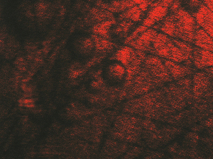
Carbon-fiber with air microspaces inside composite |
Bunch of C-fibers 1 mm under surface of the organic composite |
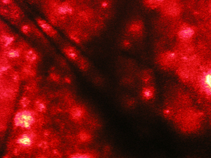
Infrared picture of structure
of the sample |
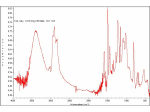
Organic composite absorption spectrum in IR |
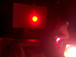
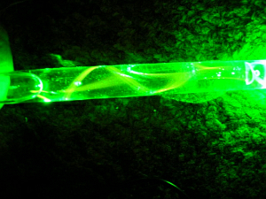
Adjustment of the laser-beam expander |
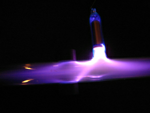
Plasma in magnetic field |
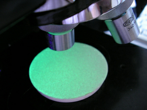
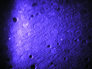
Luminescence of ZnS in UV radiation |
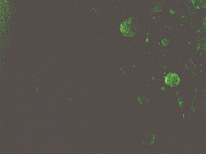
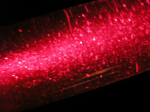
Microvacancies in the material read by microluminescent technique |
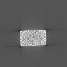
Tomogram. of structure of the material |
Polarizing microscopy in practice |
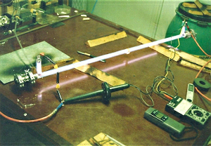
The history: making of the prototype of air-cooled CO2 laser 60 W |
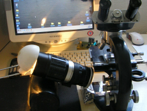
Preparation of the measuring laser |
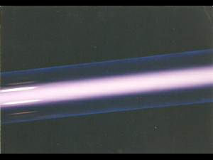
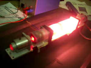
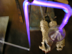
Plasma in the laser-tube |
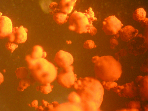
Clusters of the Fe2O3, 800x magnified |
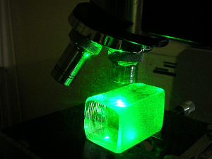
2. harmonic of the YAG-laser in microscopy |
|
The structure of the liquid
|
Organic fluid properties
|
Unconventional analysis of the compound through the gaseous discharge |